Rhinoplasty is one of the most intricate and precise cosmetic surgeries, requiring a high level of expertise and planning. In recent years, 3D imaging technology has revolutionized the way rhinoplasty is performed, improving both the precision of the surgery and the overall outcomes for patients. This cutting-edge technology allows surgeons to create detailed, virtual models of a patient’s nose, offering a better understanding of the nasal anatomy and helping patients visualize their potential results. In this article, we’ll explore the role of 3D imaging in rhinoplasty surgery in Dubai(تجميل الأنف في دبي ), its benefits, and how it is shaping the future of nasal surgery.
What is 3D Imaging in Rhinoplasty?:
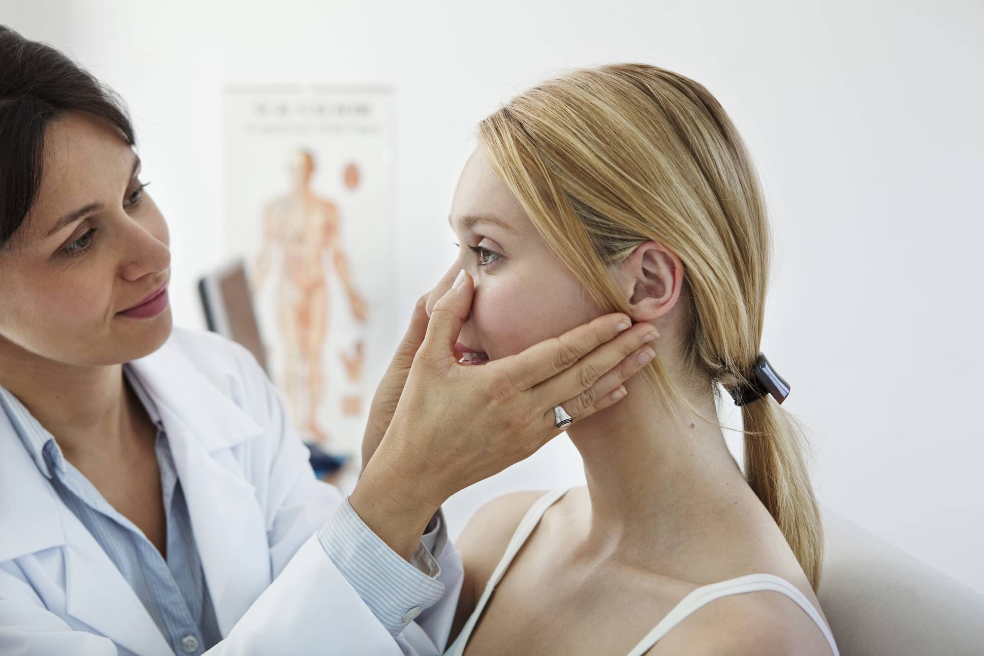
3D imaging in rhinoplasty refers to the use of specialized technology to create a three-dimensional digital representation of a patient’s nose and surrounding facial structures. This imaging allows surgeons to assess the nose from various angles and make detailed plans for the procedure.
1. How 3D Imaging Works:
Using advanced software and high-resolution cameras, a 3D scan of the patient’s face is captured, creating a virtual, lifelike model. This model is used to analyze the nose’s shape, size, and proportions in relation to the rest of the face. Surgeons can then manipulate the model to simulate different changes, such as reducing a hump or reshaping the nasal tip, allowing for a more accurate surgical plan.
2. Virtual Simulation of Results:
One of the most powerful features of 3D imaging in rhinoplasty is its ability to simulate potential outcomes. Patients can see a virtual version of their nose after surgery, giving them an idea of what their new appearance will look like. This helps to set realistic expectations and provides an opportunity for the patient and surgeon to discuss and refine the desired result before surgery.
Benefits of 3D Imaging in Rhinoplasty:
The integration of 3D imaging into rhinoplasty has offered numerous advantages, both for the surgeon and the patient. The technology enhances precision, allows for more personalized results, and improves communication throughout the surgical process.
3. Enhanced Precision and Planning:
With traditional rhinoplasty techniques, surgeons had to rely on their experience and a limited number of views to make decisions about reshaping the nose. However, 3D imaging provides a more comprehensive view of the nasal anatomy, enabling surgeons to plan the surgery with much greater precision. This detailed visualization helps minimize errors and optimizes the likelihood of achieving the desired outcome.
4. Personalized Approach:
Every patient’s facial anatomy is unique, and 3D imaging allows for a highly personalized approach to rhinoplasty. Surgeons can take into account the individual’s facial proportions and structure, ensuring that the new nose complements the rest of the face. This custom-tailored approach results in more natural-looking and harmonious results, which are more likely to meet the patient’s aesthetic goals.
5. Clearer Communication with Patients:
One of the challenges of traditional rhinoplasty consultations is conveying the possible results to patients. While a surgeon’s verbal description or a set of before-and-after photos may offer some insight, they cannot provide the same level of detail as a 3D image. The ability to show patients a visual representation of their potential results allows for clearer communication, reducing confusion and increasing satisfaction with the final outcome.
6. Improved Surgical Confidence:
Having a clear and detailed plan based on 3D imaging can increase a surgeon’s confidence in the surgery. The technology allows surgeons to see potential complications and adjust the approach accordingly. As a result, there is a lower risk of complications and the surgery can be performed with a greater sense of certainty, leading to better results for the patient.
3D Imaging for Preoperative Planning:
3D imaging plays a pivotal role in the preoperative planning phase of rhinoplasty. Surgeons use the technology to simulate various surgical techniques and assess how different changes will affect the overall appearance of the nose.
7. Assessing Nasal Symmetry:
3D imaging allows surgeons to examine nasal symmetry with remarkable precision. Many patients seek rhinoplasty to correct asymmetry or other structural issues, such as a deviated septum. Using the 3D model, surgeons can plan the necessary adjustments to achieve optimal alignment and facial harmony.
8. Predicting and Preventing Complications:
By examining the 3D model, surgeons can identify potential complications before the procedure even begins. For instance, a surgeon can assess whether there is enough cartilage for a nasal tip modification or whether there is a risk of excessive narrowing of the nostrils. This proactive approach helps to avoid common pitfalls and enhances the safety and success of the surgery.
9. Simulating Different Techniques:
There are several rhinoplasty techniques available, including open and closed rhinoplasty, each with its own advantages. With 3D imaging, surgeons can simulate different approaches and determine which one will yield the best results. Whether it’s reducing a nasal hump, refining the nasal tip, or correcting breathing issues, 3D imaging allows surgeons to explore multiple options and choose the most effective one for the patient.
3D Imaging in the Postoperative Phase:
3D imaging doesn’t just play a role in the preoperative planning; it is also beneficial in tracking a patient’s progress after surgery. Postoperative 3D scans can help surgeons monitor healing and assess the final outcome of the procedure.
10. Monitoring Healing and Adjusting Expectations:
After rhinoplasty(تجميل الأنف ), patients are eager to see the final results. However, the healing process can take several months, and swelling can obscure the true shape of the nose. By using 3D imaging, surgeons can track the progress of healing and evaluate how much swelling has subsided. This can help patients adjust their expectations and better understand the healing process.
11. Creating a Clear Roadmap for Revision Surgery:
In some cases, patients may require revision rhinoplasty to refine their results or address any complications. If a revision is necessary, 3D imaging can provide a clear roadmap for the surgeon to work from. By comparing preoperative and postoperative 3D scans, the surgeon can pinpoint areas that require adjustment and ensure the revision is carried out with precision.
How 3D Imaging Enhances Patient Satisfaction:
One of the most significant benefits of 3D imaging in rhinoplasty is the improvement in patient satisfaction. The ability to visualize potential results before surgery ensures that both the surgeon and the patient are on the same page. The technology allows for better-informed decisions and more realistic expectations, ultimately leading to a more positive surgical experience.
12. Reducing Anxiety:
Many patients experience anxiety when considering rhinoplasty, as they are uncertain about how their new nose will look. 3D imaging can reduce this anxiety by providing a realistic preview of the final result, helping patients feel more confident and comfortable moving forward with the procedure.
13. Better Post-Op Satisfaction:
Because patients can see a virtual representation of their nose before surgery, they are better equipped to understand the changes that will occur. This clarity often leads to a higher level of satisfaction after surgery, as patients have a more realistic expectation of the final outcome.
Conclusion:
3D imaging is a game-changer in the field of rhinoplasty, offering numerous advantages for both surgeons and patients. From enhanced precision in preoperative planning to improved postoperative monitoring, the role of 3D imaging has revolutionized the way rhinoplasty is performed. For patients seeking rhinoplasty, particularly in Dubai, this technology ensures a more personalized, accurate, and satisfying experience. With 3D imaging, rhinoplasty is not only safer but also more predictable, offering patients the best possible results.